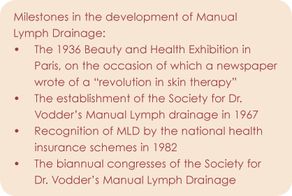
One would think that Dr. Vodder’s Manual Lymph Drainage (MLD) enjoyed a steep rise to success. After all, for whatever reasons, it did manage to survive the reefs of recession and the cost restraining law land is thus still covered by the national health insurance schemes in Germany, by which it was recognized only 25 years previously. Yet, it took half a century before it was firmly established as a technique in physical therapy.It was a long journey, beginning in 1932 when Vodder, a Danish scientist, not only discovered MLD intuitively, but also endeavored to elucidate the technique theoretically on the basis of the somewhat obscure literature on the lymph system,
which was still founded essentially on macroscopic studies.
In order to describe MLD today and reveal its underlying mechanisms of action, we draw reference to Vodder’s description of his method: The nature of the stimulation gives rise to a corresponding effect, with pressure, contact area, frequency and rapidity of the manipulations, stretching of the skin, etcetera having a variable effect.
In this respect then, MLD is a technique based on certain guidelines, but one that depends primarily on the finger
control of the therapist, who strives to exert a fluid displacing effect on both drainage systems, the venous and lymphatic, instead of a hyperemizing effect.
Body fluids can be displaced both intra-vascularly and extra-vascularly; that is to say in the vessels and in loose connective tissue. In the vessels is performed either mechanically by centripetal strokes, or by stimulation of the vascular muscles. In loose, interstitial connective tissue only mechanical displacement is possible.
 It should be recognized that owing to the function of the lymph system a certain suction effect arises, analogous to the effect of a vacuum-type water pump, which extends into the connective tissue. Proteins and protein-bound water are thus transported into the bloodstream via the lymph system. Because urinary outflow is increased in healthy subjects following MLD, it is surmised that MLD releases some water bound to mucopolysaccharides of amorphous (i.e. nonfibrillous) ground substance.
The stimulating action of MLD on the lymphatic muscles was demonstrated by Mislin in 1972. Mislin also described how the unique manipulations of MLD stimulate the lymphatic musculature: Physiologic vasomotor lymph drainage results from the autonomic pulsations of the lymphatic sections or series of lymphatic sections.
It is likely that MLD has a decisive influence on this drainage system. The process consists of rhythmically alternating phases of dilation and contraction in a successive series of metachronous activated lymph sections. This gives rise to a peristaltic wave along the lymphatic vessel. Thus, the dilation contraction frequency of the lymphatic sections are synchronized and the resulting pulsations are peristaltically metachronous. Myogenous and nervous control of vascular activities by synergistic receptors in the vessel walls ensure the coordinated transport of lymph. The main physiologic stimulation is pressure and temperature. Intravascular stretching across, but also along, the vessels increases the pulse rate of the lymphatic sections. Smooth muscle cells (e.g. those in the vessel walls) exhibit electrical and mechanical reactions upon passive stretching. Vascular muscles having autonomous (i.e. pacemaker) properties require well-dosed stretching dependent on the momentary intravascular volume in order to ensure regulation of their rhythmical repolarization that is adapted to the prevailing situation. For all these reasons MLD, which exerts a (in some respects, inadequate) physiologic tensile stimulation, stimulates the vasomotor lymph drainage system.
Extramural lymph drainage (i.e. the action of external mechanical factors on the lymphatic vessels) is founded on the fact that certain outside forces not only stimulate the vascular musculature in the manner described above but also exert a mechanical effect on the content of the lymphatic vessels. Foremost among these factors are the movements of the skeletal muscles, the pulsation of the arteries (analogous to functional coupling in the case of the veins), the peristalsis of the intestines, the movements of the diaphragm and other muscles of the respiratory system, and the variations in pressures that arise in the pectoral and abdominal cavities during respiration.
Extravascular lymph drainage involves lymph formation and extravascular circulation. The higher the content of protein in tissue, the less water can flow out of the tissue via the venous capillaries, because it is bound by protein. In assuming the function of returning protein from the tissue, the lymph system allows more water to drain from the venous capillaries.
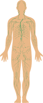
The lymph capillaries commence as blind finger-like extremities in interstitial tissue. They lack the basement membrane found in blood capillaries and consist of a single, thin layer of partially overlapping endothelial cells connected radically at one end to stretch resistant collagen fibers of the connective tissue by extremely fine precollagenous (matrix) filaments. If the connective tissue swells as a result of an increased influx of water, the pressure within the interstitium increases. As a result, the collagen fibers distend, pulling the nonelastic anchor filaments of the lymph capillaries with them. The intercellular spaces in the lymph capillaries widen, so that there is an increased flow of water, macromolecule, large and small particles (sometimes including erythrocytes) into the capillary lumen.
As the lymph capillaries fill, the pressure in the interstitial tissue decreases due to the efflux of water, and the pressure in the lymph capillaries increases due to the influx of water. These pressure changes cause the endothelial cells, which act like flutter valves, to close the intercellular gaps, so that the lymph capillaries present as filled and closed channels. The anchor filaments have also returned to their initial position, since the water content and the pressure in the connective tissue have decreased. They would undergo a relative increase if proximal emptying of the lymph capillaries (e.g. as brought about by MLD) caused the intracapillary pressure to drop. The result of such a relative increase in interstitial tissue pressure is intercellular filtration into the lymph capillaries (a further active protein-transport mechanism as transcellular cytopempsis). This is the mode of action in the lymph capillaries as described by Mislin, Foldi and Casley-Smith, and in a recent publication by Tischendorf.
In addition to the endothelial cells with their overlapping edges, there are also the usual closely spaced endothelial cells whose edges are connected by intercellular cement, especially hyaluronic acid. The protein molecules migrating in the direction of the lymph capillaries open these cemented spaces by pushing hyaluronidase in front of them. This enzyme liquefies (depolymerizes) the cement so that protein can flow through the openings into the lymph capillaries. The above described “flutter valves” with all their consequences result in proportion to the conversion of tight cell junctions to gap junctions caused, for example, by the action of hyaluronidase (i.e. The temporary liquefaction of intercellular cement). As soon as the action of hyaluronidase ceases the taught junctions are re-established, and the cycle begins anew.
G. Hauck (1980), like Casley-Smith (1976), has described variable prelymphatic tissue spaces and channels in the ground substance of connective tissue which have the function of returning proteins and fluid relatively quickly to the lymph capillaries. He maintains that the elastic fibers of the connective tissue serve as “guide rails” for the transport of fluids, since the flow rate of fluids is faster along the elastic fibers than elsewhere. These channels, which can only be seen under the electron microscope, are far smaller in diameter than the initial lymph capillaries and reveal no wall structure of their own. The beginnings of wall formation can also be marked by dark field and fluorescent microscopy and recognized as the site of continuous transition to the lymph capillaries.
These findings support the view of the lymph system as a converging drainage system open at the periphery. It may be assumed that the lymph capillaries function like suction drains. Such drains are used, for example, in agriculture where they draw water directly from the soil and deliver it to collection drains. Again, the lymph capillaries could be likened to the sap channels in plants which transport water from the soil up into the leaves and in which similar mechanisms are at work.
During MLD therapy unbound water in loose connective tissue also flows out of the interstitium at an increased rate via the venous capillaries. The result is, like that of a surgical stocking, decreased edema, provided that the fluid in question is low in protein content. Thus, MLD promotes, on the one hand, water drainage via venous capillaries and on the other hand, the removal of water and protein via the lymph system. Complications arise when the lymph system becomes incompetent, owing to the extirpation of lymph nodes or their fibrous obliteration by irradiation so that lymph drainage of whole regions is no longer ensured.
In such cases, MLD attempts to push the lymph against the normal drainage direction via the valveless lymph capillaries and the valved (but in contrast to the subsequent “transport vessels” still muscle free) “guiding vessel” as found in the superficial network of the skin lymphatics, rerouting it to regions still supplied with sufficient lymph vessels. Transport is against the normal direction of drainage, because the lymphatic vessel apparatus no longer functions due to the blockage or absence of regional lymph nodes. It is possible to transport the lymph in the skin lymph vessels deeper into the cutis or subcutis via so-called watersheds and interterritorial anastomoses. It should be kept in mind that the smallest lymphatic vessels of the skin, referred to above as “guiding vessels” and which already possess valves, can be influenced by MLD in such a way that the valves reverse position and the direction of lymph drainage can be controlled.
Furthermore, substances which must be transported via the lymph system can be pushed along in tissue spaces until they reach functional lymphatic vessels. Among the various applications of MLD, chronic cases of inflammation should not be forgotten, these falling in the domain of various disciplines. Inflammation is always accompanied by edema, which is initially situated between fibrils in loose interstitial connective tissue but later also involves the fibrillary structures themselves. When we mobilize this interstitial edema by means of MLD, we at the same time remove other lymph-borne substances that sustain the inflammation. Thus, chronic inflammation is a prime indication for MLD.
The manual pressure used in MLD depends on various factors:
- The pathologic tissue condition being treated
- The type of tissue being treated
Thus, in the case of leg ulcers or recent hematomas the pressure used is markedly different from that employed in the case of severe fibrosis. Also, the thickness and nature of the individual skin layers, especially the subcutis and the subcutaneous adipose layer are important factors, as is the turgor of the skin.
In addition, the pressure used depends to a large extent on the regionally variable substrate underlying the skin; bone, cartilage, tendon or aponeurosis, fascia and ligament. In the most frequent case, with a firm muscular substrate, muscle tone also plays a key role.
The question as to whether and to what extent the massage pressure can spread laterally also has a considerable influence on the pressure used. In the treatment of edema, the nature of the edema is of importance, namely its extent, its tension or hardness, and special properties such as its sol-gel relation, protein and fiber content, and sensitivity to pain.
Under no circumstances can an “optimal” manual drainage pressure be calculated. It can only be learned through many comparative treatments by teacher and student during MLD training and in the subsequent accumulation of experience until a feel for this difficult therapeutic form is gradually developed.
 Dr. Reinhard Bergel is the president and founder of H-e-a-t. Inc (Health-enhancement-accessories-training) Spa Kur Therapy Development. He is an advisor in Spa Kur facility development and conducts spa staff training. He has published numerous articles as well as the comprehensive SPA ENCYCLOPEDIA. Bergel is a founding member of the American Society of Lymphology and American Day Spa Association and has operated a health spa clinic and physical rehabilitation center for almost two decades. He has been practicing and teaching Lymphedema Management since 1986.
Want to read more?
Subscribe to one of our monthly plans to continue reading this article.